
Advanced 3D Dental Imaging for Precise Treatment

Advanced 3D Dental Imaging for Precise Treatment Planning and Better Patient Care
By Carmy Michael, Dental Care of Rialto
Three‑dimensional dental imaging has transformed diagnosis and planning by giving dentists clearer, more reliable information for better outcomes. Cone Beam Computed Tomography (CBCT) captures detailed 3D views of teeth, jaws, and surrounding anatomy, helping clinicians make smarter treatment choices and avoid surprises. Below, we outline key technologies, their role in treatment planning, and what patients should know about CBCT and safety.
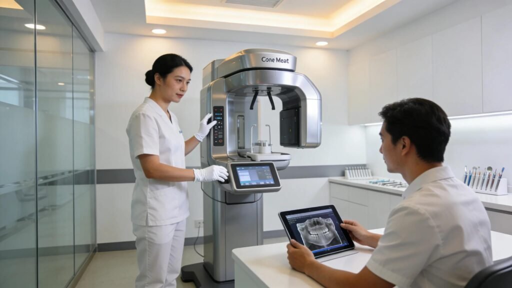
Advanced Technologies Utilized:
Dental imaging now pairs advanced scanners with intuitive software and digital workflows to produce high‑resolution 3D images. CBCT provides accurate multiplanar scans; when combined with guided‑surgery tools and cloud platforms, teams can plan and execute procedures with greater precision, reducing invasiveness and speeding recovery.
Influence on Treatment Planning:
3D scans map teeth and bone in true spatial context, eliminating the limitations of flat X‑rays. With detailed visuals, clinicians can predict outcomes, tailor plans, and reduce intraoperative guesswork—often shortening procedures and improving long‑term success.
At Dental Care of Rialto we use in‑office CBCT and 3D services to support personalized planning for implants and complex procedures, helping ensure each step is deliberate and well informed.
Improvement in Patient Care:
Advanced imaging supports minimally invasive techniques that often mean quicker recoveries and less discomfort. Clear 3D images improve communication so patients feel informed and receive more natural‑looking outcomes.
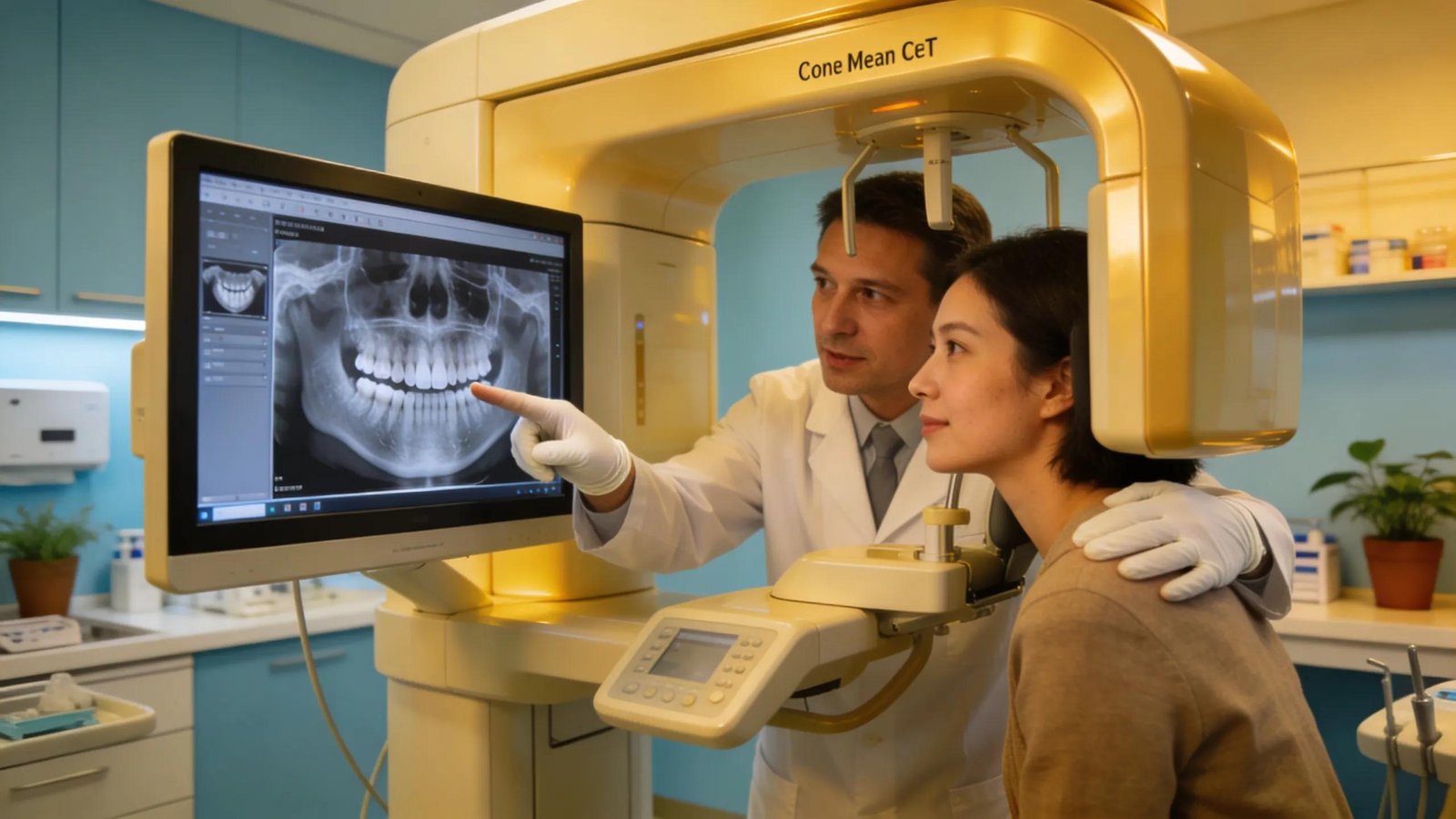
What is Cone Beam CT and How Does It Enhance 3D Dental Imaging?
Cone Beam CT (CBCT) produces volumetric scans that reveal depth and spatial relationships, providing critical detail for diagnosis, planning, and surgical guidance beyond 2D X‑rays.
How Cone Beam CT Technology Works in Dental Diagnostics
CBCT rotates a cone‑shaped X‑ray beam around the patient to collect multiple images; software reconstructs these into a 3D model that highlights bone contours, nerve paths, and sinus anatomy for accurate assessment.
Comparison Between CBCT and Traditional Dental X-Rays
CBCT offers true 3D views without overlapping shadows of 2D films, giving clearer insight into complex anatomy. It often provides needed diagnostic detail at radiation levels appropriate for dental care.
This comparison shows how CBCT provides clearer, actionable information for safer, more predictable care.
How Does Advanced 3D Dental Imaging Improve Precision in Dental Treatments?
3D imaging supplies fine anatomical detail—bone density, nerve location—that enables guided surgical techniques and greater accuracy, reducing complication risk and improving functional and aesthetic results.
Benefits of 3D Imaging for Accurate Dental Implant Placement
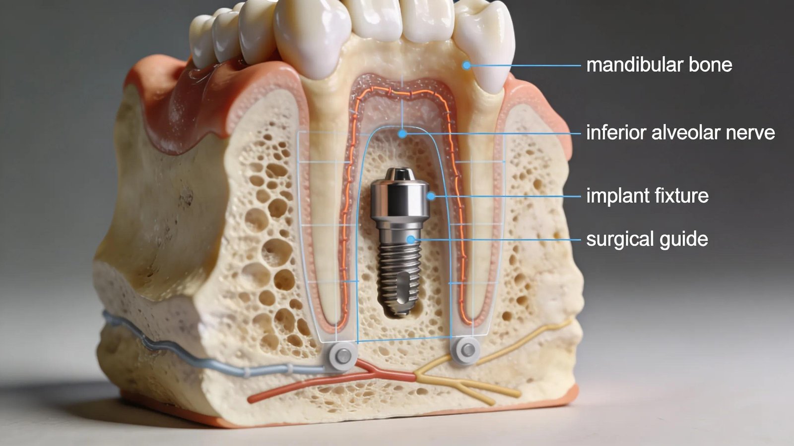
For implants, 3D imaging helps select implant size, angle, and position while avoiding vital structures, increasing long‑term success and reducing corrective procedures.
These differences explain why clinicians prefer 3D imaging for implants and advanced treatments.
Role of Digital Dental Diagnostics in Reducing Treatment Complications
Integrating scans with planning software and guided tools lets teams anticipate challenges and adjust plans, preventing problems and supporting safer, more efficient care.
What Are the Safety Considerations and Radiation Exposure Levels in 3D Dental Imaging?
Safety is essential: clinicians use the lowest reasonable dose for diagnostic quality and follow protocols to protect patients, ensuring benefits outweigh risks.
Radiation Dose Ranges for Cone Beam CT Scans Compared to Other Imaging
Dental CBCT balances image quality with dose reduction; in many cases it exposes patients to less radiation than larger medical CTs while delivering necessary detail.
Patient Preparation and Safety Protocols for CBCT Imaging Procedures
Preparation typically includes a brief medical history review, removing jewelry or metal, and staff instructions. For pregnancy or special concerns, clinicians discuss alternatives and precautions.
Why Should Rialto Patients Choose Local Advanced 3D Imaging Services?
Local access to CBCT makes care more convenient and timely for Rialto residents, reducing travel and enabling faster, coordinated treatment for complex cases.
Access to Cutting-Edge Cone Beam CT Technology at Dental Care of Rialto
Dental Care of Rialto offers in‑office CBCT so patients receive precise diagnostics without referrals, reflecting our commitment to accurate, patient‑centered care.
Patient Success Stories Demonstrating Improved Outcomes With 3D Imaging
Patient stories demonstrate shorter recoveries, clearer expectations, and better long‑term results when 3D imaging guides treatment.
Frequently Asked Questions
What are the advantages of using advanced dental imaging over traditional methods?
3D imaging reveals hidden anatomy for more accurate diagnoses and tailored plans, often enabling less invasive procedures and efficient exams.
Are there specific dental conditions that benefit most from 3D imaging?
Implants, impacted teeth, jaw pathology, orthodontics, and complex surgical cases benefit most from 3D imaging due to bone and nerve visualization.
How does digital dental diagnostics integrate with patient care?
Scans feed planning software, guide surgical templates, and improve clinician–patient communication, shortening appointments and clarifying expectations.
What training do dental professionals need to utilize 3D imaging technology?
Clinicians need training in acquisition and 3D dataset interpretation; many dental programs and continuing education courses cover CBCT use.
Is there a risk of radiation exposure with Cone Beam CT scans?
CBCT involves radiation, but units are optimized to use the lowest necessary dose; dentists follow protocols to limit exposure when benefits are justified.
How do patients prepare for a Cone Beam CT scan?
Expect to remove metal accessories and share medical history; if pregnant or concerned, inform your team so they can advise the safest approach.
Conclusion
Advanced 3D dental imaging brings clarity and confidence to treatment planning, supporting safer procedures and better outcomes. At Dental Care of Rialto, our modern imaging tools are part of delivering thoughtful, effective dental care—ask us how 3D imaging can improve your treatment.