
Why Dental X-rays Are Crucial for Your Oral Health
The Importance and Benefits of Dental X-Rays
By Michael Carmy, Dental Care of Rialto
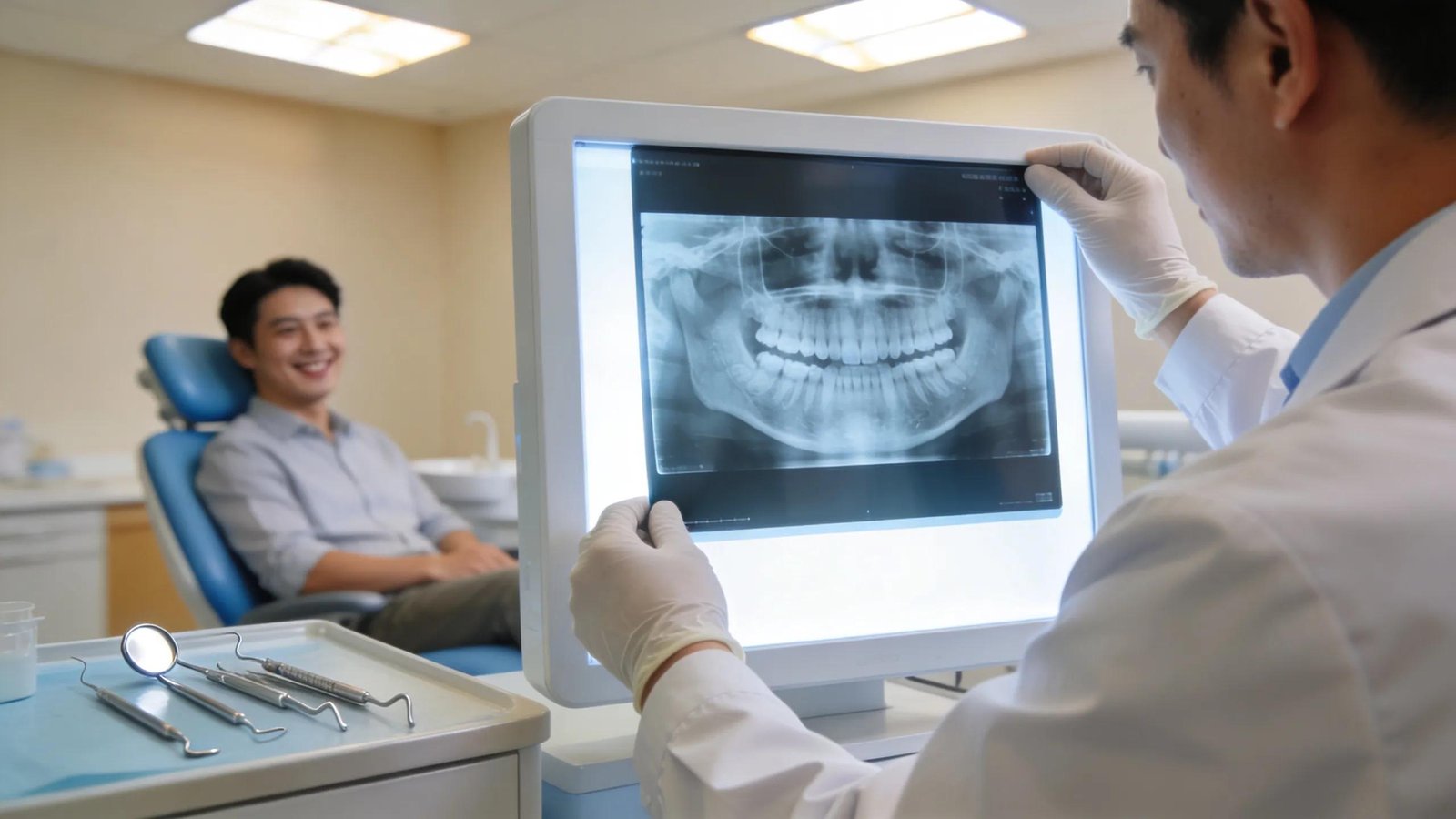
Dental X-rays are a key diagnostic tool in modern dentistry. They reveal conditions hidden in a clinical exam—such as interproximal decay, periodontal bone loss, and infections—and guide treatment planning. This article describes common X-ray types, their diagnostic roles, implant planning use, and safety practices at Dental Care of Rialto in Rialto, CA.
What Are the Main Types of Dental X Rays Used in Oral Health Diagnostics?
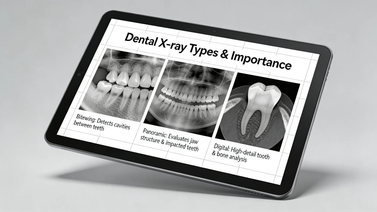
Dental teams select specific X-ray formats to visualise structures and detect pathology not evident on inspection.
- Bitewing X-rays: Detect cavities between teeth by imaging upper and lower crowns together.
- Panoramic X-rays: Give a broad view of jaws, teeth, and supporting structures for overall assessment and bone evaluation.
- Digital X-rays: Use lower radiation, deliver instant images, and simplify storage and sharing.
Together these modalities enable a more complete oral health assessment.
How Do Dental X Rays Detect Hidden Dental Problems Effectively?
X-rays identify problems early, reducing the need for more invasive care later.
Cavities that start between teeth or beneath restorations are often invisible on exam; radiographs reveal interproximal decay and issues under fillings and allow assessment of supporting bone for early periodontal disease.
Research evidence supports the diagnostic value of radiography in detecting important dental conditions.
Accuracy of Radiography for Proximal Caries Detection
Proximal caries detection accuracy using intraoral bitewing radiography, extraoral bitewing radiography and panoramic radiography — K. Kamburoğlu, 2012
Beyond cavities, X-rays show bone loss and root infections such as periapical abscesses, enabling timely diagnosis and treatment planning. Routine radiographic review supports prompt clinical intervention.
Dental Care of Rialto emphasises radiography as part of comprehensive, evidence-based care.
What Types of Issues Identified:
Dental X-rays commonly reveal:
- Cavities: Early interproximal or sub-restoration decay.
- Gum Disease: Bone levels and structural changes indicating periodontal disease.
- Infections: Abscesses, cysts, and other pathoses not detectable visually.
These findings explain why targeted radiographs are often part of routine exams.
The use of advanced tools, including deep learning, is improving radiographic interpretation and detection accuracy.
Deep Learning for Caries Lesion Diagnosis in Bitewing Radiographs
This study evaluated the effectiveness of deep convolutional neural networks for diagnosing interproximal caries in digital bitewing radiographs — Y. Bayraktar, 2022
What Measures Ensure Radiation Safety During Dental X Ray Procedures?
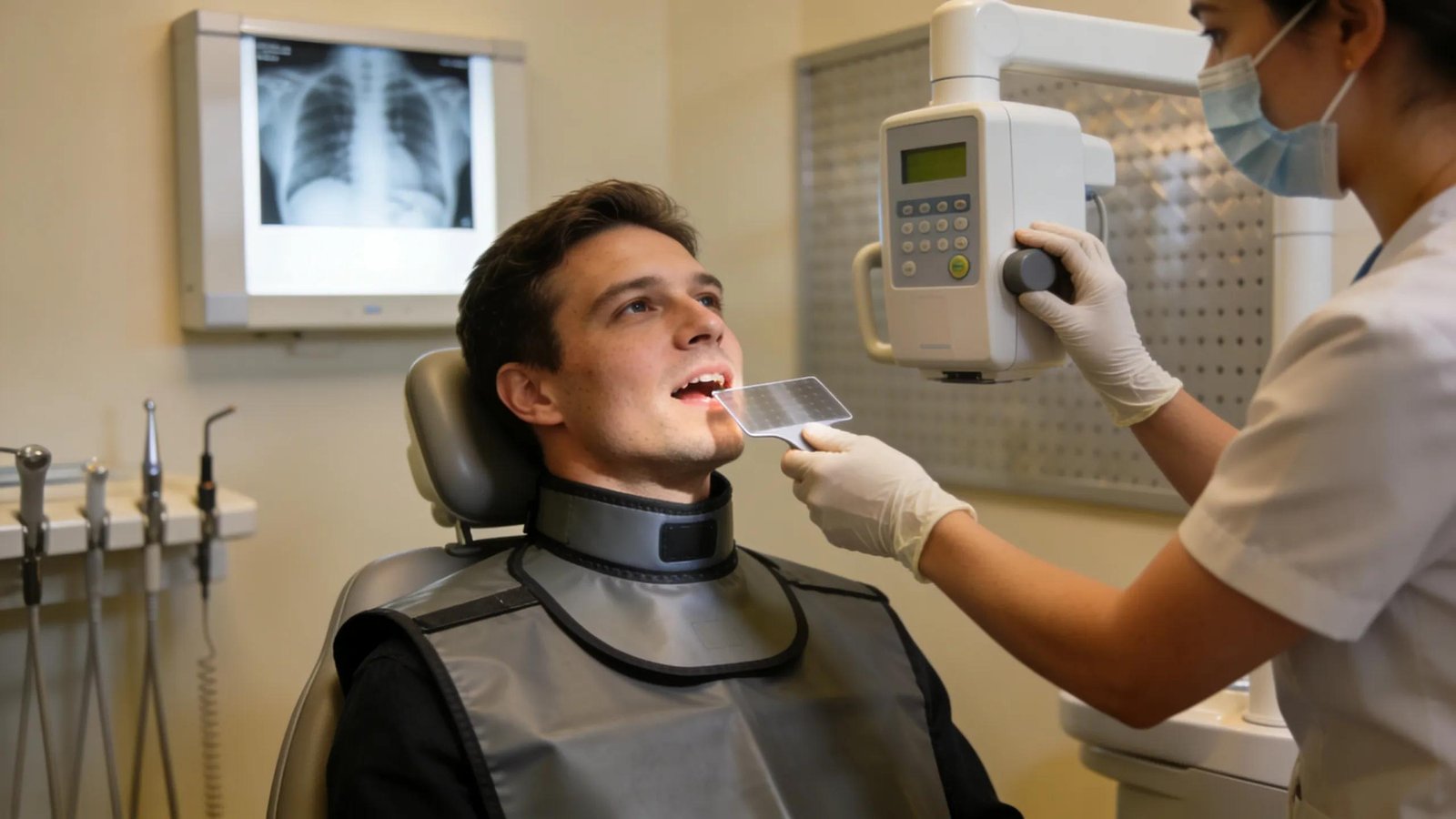
Radiation safety is fundamental; standard protocols minimise exposure during imaging.
- Lead Aprons and Collars: Shield sensitive areas during exposures.
- Digital X-ray Technology: Requires less radiation than film and reduces repeat images.
- Precise Targeting: Focused beams and collimation limit dose to adjacent tissues.
Applied together, these measures provide necessary diagnostic information with minimal risk.
How Do Dental X Rays Support Treatment Planning, Including Dental Implants?
X-rays give objective imaging that informs clinical decisions and sequence of care tailored to each patient.
For implants, radiographic assessment evaluates bone volume and anatomy to plan implant position and angulation accurately.
Contemporary analytic tools further streamline interpretation for treatment planning.
Deep Learning Enhances Dental X-ray Diagnostics
Applying deep learning techniques to dental X-ray images can assist clinicians in detecting conditions such as cavities, restorations, and implant-related findings more efficiently — Z. Ali, 2024
Careful radiographic analysis is integral to planning at Dental Care of Rialto and helps optimise outcomes.
What Role Do X Rays Play in Planning Dental Implant Procedures?
When planning implants, X-rays provide essential information including:
- Bone Density and Volume: Assess quantity and quality of supporting bone.
- Identification of Complications: Reveal anatomical variations or obstacles that could affect surgery.
These data help clinicians form a clear surgical plan and anticipate adjunct procedures if needed.
How Do Dental X Rays Integrate With Other Oral Diagnostic Services?
Radiographs complement clinical exams and other modalities to produce a complete diagnostic picture.
- Comprehensive Assessments: Combining clinical findings and imaging clarifies diagnosis.
- Guidance for Further Testing: X-rays indicate when CT scans or specialist assessments are warranted.
- Enhanced Patient Communication: Images help patients understand conditions and treatment plans.
This integration reinforces the role of X-rays in evidence-based dental care.
Understanding each X-ray’s role helps patients appreciate their value in prevention, diagnosis, and planning.
Frequently Asked Questions
What should I expect during a dental X-ray procedure?
You will sit while the clinician positions the device and places a lead apron. For intraoral images you may bite a short holder; the process takes only a few minutes and is typically comfortable.
How often should I have dental X-rays taken?
Frequency depends on your oral health and risk profile. Many adults have X-rays every one to two years; those with active issues or treatment needs may require imaging more often. Your dentist will advise a schedule based on clinical findings.
Are there any risks associated with dental X-rays?
Modern dental X-rays use low radiation. Digital systems and protective measures (aprons, collimation) minimise exposure; the risk is considered minimal when protocols are followed.
Can dental X-rays detect problems other than cavities?
Yes. Radiographs reveal periodontal bone loss, abscesses, impacted or unerupted teeth, and structural jaw issues, and they help evaluate existing restorations.
How do dental X-rays improve treatment outcomes?
They enable early detection and precise planning—for example, assessing bone for implants improves placement accuracy and long-term success.
What alternatives to dental X-rays exist for diagnosing oral health issues?
Alternatives include clinical examination and, when needed, advanced imaging like CT scans for three-dimensional detail. CTs are used selectively due to cost and higher radiation; routine care relies on conventional X-rays.
Conclusion
Dental X-rays are essential for finding hidden conditions and planning effective, personalised treatment. By revealing cavities, bone changes, and infections early, radiography enhances patient safety and treatment success. Contact Dental Care of Rialto to schedule an appointment and receive care supported by precise diagnostic imaging.